Health Problems found in the Northern Inuit Dog
“
|
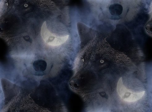
I have become increasingly concerned about the rapid decline in the health of the 'Northern Inuit type dog' in recent years. Recurring problems include hip dysplasia, elbow deformities, epilepsy, Addison's disease, von Willebrands disease, dwarfism, monorchidsm, sterile bitches, OCD, DM, overshot jaws, cataracts, organ failure (kidney, liver and heart) and various forms of cancer (lymphoma, bone cancer and brain tumours). (Proof of these health problems occurring in Northern Inuit dogs is available to visitors on request) Thankfully I have only experienced three of these problems in my lines during the time I have been breeding these dogs, namely monorchid male puppies, epilepsy from one mating that I did over ten years ago and one pup with cataracts (possibly due to the mother having had a virus while pregnant and also the pup needing to be hand reared, which can lead to eye problems, however I am not taking any chances and will not be breeding from that bitch again).
It is apparent to me, after discussing the genetic health problems with
the top experts in the country, that these ailments are compounded by the
chronic inbreeding within the breed causing gene mutations, also insufficient care being taken in
the selection of the founder dogs. The lack of genetic
diversity means the future does not look promising and new blood is
desperately needed. In this respect myself and a few responsible breeders have decided to bring in new blood and create our own unique, healthier lines of wolf-lookalike dogs, which will be registered as part of the Anglo Wulfdog breeding programme. Contrary to claims on some Northern Inuit dog websites we are not passing our dogs off as Northern Inuit dogs, but are in fact doing the opposite by making the point of distinguishing our dogs from Northern Inuit dogs by calling our dogs "Anglo Wulfdogs". We do not wish to be associated with Northern Inuit or any other wolf lookalike dog in any way. For more information on all these health issues please refer to the Health section of my forum, where they are dealt with in more detail. If anyone owns, or has owned in the past, a 'Northern Inuit type dog' with any health issues I will be pleased to hear from you as it would assist with my research into the health of the breed. There is an easy to complete online survey form available on my forum and on the health section of this website. All information will be kept in the strictest confidence and used solely to benefit future breeding of this type of dog. |
|
Monorchidism & Cryptorchidism Monorchidism literally means one descended testis, and is very rare in most breeds but quite prolific in the Northern Inuit dog.
Cryptorchidism is the retention of one or
both of the testes, usually in the abdomen. Cryptorchid testes can also
be outside the body wall under the skin. The danger with cryptorchidism
is that if the testis is in the abdomen, it is being exposed to a higher
body temperature and is at great risk for Sertoli cell tumor (13 times
more often than dogs with descended testes). Affected animals should be
castrated (neutered). It is suspected to be a hereditary condition. Dogs
which are unilateral cryptorchid (one testis is descended, one is not)
can still be fertile and can pass the trait on to offspring.
Cryptorchidism occurs most frequently in purebred dogs, in certain
breeds, and within certain families of a breed.
|
| Inherited Diseases of the Eye
(not
all are found in the NI and eye problems are generally rare) Retinal Dysplasia describes those inherited conditions in which abnormal differentiation results in neuroretinal fold and rosette formation, degeneration or non-attachment. The effect ranges from no noticeable impairment of sight through to blindness. All forms of RD are considered simple recessive traits. Collie Eye Anomaly enjoys high incidence in the Rough Collie and Shetland Sheepdog breeds in the U.K, but is also seen in the Smooth Collie and the Border Collie. It was recently recorded in a non-collie breed, the Lancashire Heeler. Again, the disease is considered to be inherited as a simple recessive trait Persistent Hyperplastic Primary vitreous is due to the retention of elements of the foetal vascular supply to the lens, the tunica vasculosis lentis. The lesions seen are variable amounts of fibrovascular plaque on the posterior lens capsule and possible posterior cortical cataract. The effect on sight can range from nothing to blindness. Removal of the diseased lens can be complicated by any vascular involvement Hereditary Cataract may be congenitally present as a nuclear opacity or may develop variably in terms of position, extent and age in both young and ageing adults from approximately 6–8 months to nine years of age. Cataract is defined as any opacity of the lens and/or its capsule. Thus, the clinical features seen range from pinhead marks to total lens opacity. The larger the cataract, the more severe the effect on sight. Congenital cataract is usually static, allowing vision through the adult cortical portion of the lens Lens luxation is the displacement or dislocation of the lens from its position on the anterior face of the vitreous due to the degeneration of its suspensory apparatus. In most patients, the lens moves into the pupil or the anterior chamber resulting in secondary glaucoma. Lens luxation is recessively inherited Progressive Retinal Atrophy covers a number of inherited neuroretinal degenerations, some of which are due to primary photoreceptor dysplasia, whilst others are due to photoreceptor degeneration of, as yet, undetermined aetiologies. Currently, fifteen breeds are involved in the PRA story in the United Kingdom and its possible presence is being investigated in another six breeds. All types of PRA are inherited as recessive traits and all are characterised ophthalmoscopically by increased tapetal reflectivity and blood vessel degeneration. The age of onset varies, but total blindness is the common endpoint. Retinal Pigment Epithelial Dystrophy is due to an inability of the retinal pigment epithelial cells to supply nutrients to the neuroretina and as such, there ensues secondary photoreceptor degeneration. Its effect is largely confined to the non-pigmented part of the retina within the tapetal fundus and, as such, affected dogs rarely lose their peripheral vision. Primary Glaucoma is due to inherent defect within the aqueous drainage pathway. Open angle glaucoma is uncommon in the canine population, but an angle closure glaucoma, consequent to a goniodysgenesis in which there is abnormal differentiation of the pectinate ligament, is inherited in several breeds of dog. This latter disease is characterised by sudden onset loss of sight and pain due to spontaneous closure of the ciliary cleft. The goniodysgenesis can be detected at four to six months of age by gonioscopy and the disease has been described in the Basset Hound, Flat Coated Retriever, Siberian Husky, American Cocker Spaniel, Cocker Spaniel, and Welsh Springer Spaniel in the United Kingdom. Its possible presence as an inherited defect. |
| Cataracts What is a Cataract? Like a camera, eyes have a clear lens inside them that is used for focusing. A cataract is an opacity within the lens. The opacity can be very small (incipient cataract) and not interfere with vision. It can involve more of the lens (immature cataract) and cause blurred vision. Eventually the entire lens can become cloudy and all functional vision lost. This is called a mature cataract. Some mature cataracts will transform over time into hypermature cataracts. Hypermature cataracts usually are reduced in size due to loss of water and proteins from the lens. This causes the lens to shrivel and the lens capsule to wrinkle - similar to a grape turning into a raisin. Hypermature cataracts vary in how cloudy they are. Some are completely cloudy and others have clear areas that can allow some vision IF the rest of the eye is functional. Depending on the dog's age and breed, it can take several months to years for a mature cataract to turn into a hypermature cataract. Causes of Cataracts Cataracts have many causes in dogs and sometimes it is not possible to identify the cause of cataracts in affected patients. Most cataracts in dogs are inherited and can occur at any age. The cataract may develop rapidly over weeks, or slowly over years and occur in one or both eyes. Different breeds of dogs have different characteristics of cataract development. For example, cataracts in Bicho Frise dogs tend to develop rapidly in early adulthood and usually involve the entire lens in both eyes. Mixed breed dogs can also develop inherited cataracts. The second most common cause of cataracts in dogs is diabetes (diabetes mellitus). 75% of diabetic dogs will develop blinding cataracts within the first year of being diabetic. Often the cataracts form very shortly after the dog becomes diabetic. Diabetic cataracts develop VERY fast - often overnight - in dogs and they are a medical and surgical emergency. The third most common cause of cataracts in dogs is toxic reaction in the lens - the lens is "sick" due to some other ocular disease or (much less commonly) due to a drug reaction. These are called "toxic cataracts". Toxic cataracts caused by ocular disease are quite common in dogs and are caused by 1. Retinal degeneration, especially Progresssive Retinal Atrophy (PRA) 2. Uveitis (intraocular inflammation) of any cause, including trauma 3. Secondary to glaucoma (increased intraocular pressure) of any cause A special type of cataract occurs in dogs in which the lens capsule is ruptured due to trauma. The trauma can be penetrating (such as a cat claw injury or pellet gun injury) or a severe blow to the eye that results in lens capsular rupture. The lens contents leak out through the hole in the capsule and cause both cataract and a severe immune-mediated reactive uveitis; the uveitis does not usually "peak" in severity until 2-3 weeks AFTER the injury occurred. It is not always apparent that the lens capsule has ruptured; often by the time this is diagnosed it is too late to save the eye and the eye needs to be removed. Thus, it is prudent to seek immediate medical attention for ANY injury to your dog's eye. Lens capsules can also rupture if the lens swells, causing the capsule to stretch and split open. This can happen in diabetic dogs and in some types of inherited cataracts that rapidly form. Cataracts can also develop due to nutritional deficiences in dogs, such as orphan puppies on an artificial milk replacer diet. These are called nutritional cataracts and they often will improve as the puppy matures. Dogs also can develop cataracts with age (often after 8 years of life). However, age related cataracts in dogs are usually small and do not significantly interfere with vision. There are many other potential causes of cataracts in dogs, such as birth defects, radiation (usually from prolonged radiation therapy for cancer of the head), infection, etc. but discussion of these causes is beyond the scope of this article. |
|
Epilepsy There are many causes of seizures but the absence of an identifiable cause is what characterises what is known as “idiopathic” or “primary” epilepsy. There is no definitive test for epilepsy and any diagnosis is tentative. One seizure alone does not result in a diagnosis of epilepsy. The seizures must be recurrent for this diagnosis to be at all reliable and it is unlikely that treatment for epilepsy would be recommended until at least two episodes had occurred. Some breeds seem to be predisposed to canine epilepsy, among them, the Belgian Terveuren, German Shepherd, Golden Retriever and Border Collie. It has been suggested that between 1 – 6% of pure bred dogs have a seizure problem Most instances of idiopathic epilepsy initially occur when the dog is between 1 and 5 years of age although it is increasingly common for dogs as young as 6 months to be diagnosed with primary epilepsy. Dogs that start fitting after the age of 5 years will almost certainly have a secondary condition causing the fitting. Types of seizure Generalised seizure: Tonic-clonic (Grand mal or mild) In the Grand mal seizure, the tonic phase desribes the animal falling and losing consciousness. This can last for 10-30 seconds before the clonic stage which involve the paddling of the limbs and chewing/champing. Other signs include salivation, urination and defecation. Petit mal seizure: A brief duration of unconsciousness, the animal may become floppy and may stare blankly. Also known as Absence Seizure. Partial seizure: also known as focal seizures, the movements are restricted to one area of the body, e.g. one limb, turning head or body to one side, facial twitches. Partial seizures can progress into a generalised tonic clonic seizure. Complex partial seizure: seizures are linked with bizarre behaviour repeated during each seizure. Examples include chewing, fly-biting, aggression, hysterical running etc. Although the animal may not lose consciousness, there may be a lack of awareness which may last minutes or hours. Cluster seizure: several seizures within a 24 hour period with periods of consciousness (however brief) in between. Status epilepticus: a life threatening condition which involves one continuous seizure of 30 minutes or more, or several consecutive seizures with no periods of normal consciousness in between. Veterinary intervention is required immediately. Breeding The issues surrounding epilepsy and breeding are complex and emotive. The lack of a test for the condition and fact that epilepsy can be carried without the carrier becoming affected, together with late onset of seizures can all make the decision whether to exclude a particular dog, parents or littermates from a breeding program a difficult call. Only in the most limited of gene pools should the use of a dog immediately related to a dog with epilepsy be considered for breeding purposes - it should be remembered that the removal of such dogs from the gene pool would further reduce the gene pool with consequential adverse affects. Symptoms, diagnosis and treatment are covered in the relevant section of my forum. |
|
Addison's disease (Hypoadrenocorticism)
Addison's disease is also known as hypoadrenocorticism. It is an
insufficient production of adrenal hormones by the adrenal gland. Since
these hormones are essential for life, this is an extremely serious
disease and it must be treated as such.
Most dogs with Addison's disease initially have gastrointestinal
disturbances like vomiting. Lethargy it also a common early sign. Poor
appetite can occur as well. These are pretty vague signs and it is
extremely easy to miss this disease. More severe signs occur when a dog
with hypoadrenocorticism is stressed or when potassium levels get high
enough to interfere with heart function. Dogs with this problem will
sometimes suffer severe shock symptoms when stressed, which can lead to
a rapid death. When potassium levels get high heart arrythmias occur or
even heart stoppage which also is fatal. In some cases, especially
secondary Addison's disease, there are no detectable electrolyte
changes.
|
| Von Willebrand's
Disease Von Willebrand's Disease is a common inherited bleeding disorder. Clotting is a complex mechanism. In addition to platelets, clot formation is the result of a long chain of chemical reactions carried out by individual molecules called 'clotting factors.' Each factor is numbered such that factor I leads to a reaction with factor II forming a new substance. This then reacts with factor III and so on to factor XII. In Von Willebrand's Disease, the dog is missing a substance, which helps the platelets form clots and stabilizes Factor VIII in the clotting process. This substance is called 'Von Willebrand's factor.' Because of the deficient clotting of blood, dogs with Von Willebrand's disease have excessive bleeding upon injury. This would be similar to hemophilia in humans. Certain breeds have a higher incidence of vWD than others. German Shepherds, Doberman Pinschers, Shetland Sheepdogs, Chesapeake Bay Retrievers, German Shorthaired Pointers, Golden Retrievers, Standard Poodles, and Scottish Terriers all have a higher than normal incidence, showing that it can be inherited. What are the symptoms? Excessive bleeding is the main symptom. Bleeding generally occurs after a wound or surgery. In these cases, the blood simply does not clot in the normal time, and bleeding is extensive. Dog's with Von Willebrand's disease may also develop nosebleeds, or bleeding from the gums. Bleeding may also occur in the stomach or intestine in which case the stool may either have blood in it, or be black and tarry. Some dogs will have blood in their urine. Bleeding into the joints also occurs, which can cause symptoms similar to those of arthritis. The diagnosis of Von Willebrand's is made through a test, which checks for the level of Von Willebrand's factor in the blood. What are the risks? These dogs, without treatment, can bleed to death following surgery, or what might be normally considered less than life threatening injuries. What is the management? Transfusions with blood collected from normal dogs is the only proven way to treat Von Willebrand's disease. Some dogs with Von Willebrand's disease also are hypothyroid - meaning they have lower than normal levels of thyroid hormone. These dogs will benefit from thyroid hormone replacement therapy. Some studies have been done which suggest a drug called desmopressin acetate (DDAVP) may help dogs with a bleeding episode. The drug can be administered intranasally (into the nose) to increase clotting. There is still some controversy over whether this treatment is effective. There is no cure for Von Willebrand's disease. Prevention through eliminating affected individuals from any breeding program is the goal of veterinary medicine today. Tests are available to determine which dogs may have this trait. All individuals with a history of this disorder in their backgrounds should be tested.
|
|
Congenital Heart Defects
Congenital defects are those that have been present since birth.
Thankfully, they are comparatively rare, accounting for only 5% of the
cases seen by vets. These will usually cause the blood flow
through the heart to become turbulent; making a distinctive whooshing
noise that vets can hear using a stethoscope. That's what is meant by a
'heart murmur'. However, if your vet tells you they've detected a heart
murmur in your dog, it's not necessarily cause for concern.
Many puppies are born with a slight heart murmur (or puppy murmur) that
clears up by itself after 4-6 months. In many cases, vets will simply
recommend a later checkup, just to be sure that the condition has
resolved itself.
Pronounced heart murmur is quite rare, but may be indicative of a
serious congenital defect. However, without specialist experience and
equipment, it can be difficult for a GP vet to know what defect is
causing the murmur. For this reason, if the murmur is pronounced, or
persists beyond puppyhood, patients will often be referred to a
specialist cardiologist.
Patent Ductus Arteriosus: a blood vessel used to bypass the lungs of pups as they develop in the womb fails to close after birth Pulmonic Stenosis: the flow of blood from the right heart to the lungs is hampered or blocked. Aortic Stenosis: the flow of blood from the left heart to the body is hampered or blocked. Hole in the Heart: a hole between the pumping chambers of the heart |
|
Hip Dysplasia
There are several contributing factors with HD - Genetics, Diet, Exercise and Environment
To understand what hip dysplasia really is we must have a basic
understanding of the joint that is being affected. The hip joint forms
the attachment of the hind leg to the body and is a ball and socket
joint. The ball portion is the head of the femur while the socket (acetabulum)
is located on the pelvis. In a normal joint the ball rotates freely
within the socket. To facilitate movement the bones are shaped to
perfectly match each other, with the socket surrounding the ball. To
strengthen the joint, the two bones are held together by a ligament. The
ligament attaches the femoral head directly to the acetabulum. Also, the
joint capsule, which is a very strong band of connective tissue,
encircles the two bones adding further stability. The area where the
bones actually touch each other is called the articular surface. It is
perfectly smooth and cushioned with a layer of spongy cartilage. In the
normal dog, all of these factors work together to cause the joint to
function smoothly and with stability.
Hip dysplasia results from the abnormal development of the hip joint in
the young dog. It may or may not be bilateral, affecting both right and
left sides. It is brought about by the laxity of the muscles, connective
tissue, and ligaments that should support the joint. Most dysplastic
dogs are born with normal hips but due to genetic and possibly other
factors, the soft tissues that surround the joint start to develop
abnormally as the puppy grows. The most important part of these changes
is that the bones are not held in place but actually move apart. The
joint capsule and the ligament between the two bones stretch, adding
further instability to the joint. As this happens, the articular
surfaces of the two bones lose contact with each other. This separation
of the two bones within a joint is called subluxation, and this causes
the resulting problems we associate with the disease.
Dogs of all ages are subject to the symptoms of hip dysplasia and the
resultant osteoarthritis. In severe cases, puppies as young as five
months will begin to show pain and discomfort during and after vigorous
exercise. The condition will worsen until even normal daily activities
are painful. Without intervention, these dogs may be unable to walk at
all by several years of age. In most cases, however, the symptoms do not
begin to show until the middle or later years in the dog's life.
Genetics:
Almost all researchers agree that there is a genetic link involved. If a
parent has hip dysplasia, then the offspring are at greater risk for
developing hip dysplasia. Some researchers feel that genetics are the
only factor involved, where others feel that genetics contribute less
than 25% to the development of the disease. The truth probably lies in
the middle. If there are no
carriers
of hip dysplasia in a dog's lineage, then he will not contract the
disease. If there are genetic carriers, then he may contract the
disease. We can greatly reduce the incidence of hip dysplasia through
selective breeding. We can also increase the incidence through
selectively breeding. We cannot, however, completely reproduce the
disease through selective breeding. In other words, if you breed two
dysplastic dogs, the offspring are much more likely to develop the
disease but will not all have the same level of symptoms or even
necessarily show any symptoms. The offspring from these dogs will,
however, be carriers and the disease may show up in their offspring in
later generations. This is why it can be difficult to eradicate the
disease from a breed or specific line.
Nutrition:
Experimentally, we can increase the severity of the disease in
genetically susceptible animals in a number of ways. One of them is
through obesity. It stands to reason that carrying around extra weight
will exacerbate degeneration of the joint in a dog with a loose hip.
Overweight dogs are therefore at a much higher risk. Another factor that
may increase the incidence is rapid growth in a puppy during the ages
from three to ten months. Experimentally, the incidence has been
increased in genetically susceptible dogs when they are given free
choice food. In a study, Labrador Retriever puppies fed free choice for
three years had a much higher incidence of hip dysplasia than their
littermates who were fed the same diet but in an amount that was 25%
less than that fed to the dysplastic group.
There have also been studies looking into protein and calcium levels and
their relationship to hip dysplasia. Both of these studies were able to
increase the level of hip dysplasia by feeding increased amounts of
calcium and protein. In the studies, though, puppies were fed greatly
increased amounts over normal recommended values and compared to animals
fed decreased amounts. They failed to compare puppies fed a normal
amount of food that had the recommended amount of protein, fat, and
calcium to those fed a diet with slightly less protein, fat, and calcium
(similar to a 'large breed puppy food').
Exercise:
Exercise may be another risk factor. It appears that dogs that are
genetically susceptible to the disease may have an increased incidence
of disease if they over-exercised at a young age. But at the same time,
we know that dogs with large and prominent leg muscle mass are less
likely to contract the disease than dogs with small muscle mass. So
exercising and maintaining good muscle mass may actually decrease the
incidence of the disease. Moderate exercise that strengthens the gluteal
muscles, such as running and swimming, is probably a good idea. Whereas,
activities that apply a lot of force to the joint are contraindicated.
An example would be jumping activities such as playing Frisbee.
|
|
Intussusceptions
Dog intussusceptions are a greatly painful condition for your dog to face and can be greatly confused with many other conditions because of the common symptoms of diarrhoea and vomiting. The problem occurs in the intestines of the dog because, like the whole digestive system the intestines move the food along by a series of contractions much like the way a worm moves, and if this motion is too violent and aggressive then it is possible that one of the sections will overlap another causing a pocket where food may get caught, thus causing pain. In most cases this condition happens because of
another problem causing diarrhoea or vomiting, which can often cause very
violent diarrhoea or vomiting which is what causes the violent
contractions of the muscles in the intestines when the dog strains. From
this the sections overlap each other and more and more waste will get
stuck in the pocket produced, making it grow and stretch longer and
become painful. When this condition is then viewed by a professional vet they will
normally be advised to have the pet undertake surgery where they will
either pull the different sections apart from each other if the problem
is in its infancy, or if the problem is far beyond normal and simple
repair then the section that is causing the problem may be cut out and
joined back together to resume normal life after the recovery period.
|
|
Lymphoma
The “typical” canine lymphoma patient is a middle aged dog presented to
the veterinarian because one or more lumps have been found. The
veterinarian rapidly determines that all of the peripheral lymph nodes
(those near the skin surface) are enlarged and firm. Usually the dog has
not been showing any signs of illness. The next step is a blood
panel and urinalysis to more completely assess the patient’s health and
one or more lymph nodes are aspirated or biopsied to confirm the
diagnosis of lymphoma. Thee average life expectancy for a patient with untreated lymphoma is about 2 months from the time of diagnosis. There are many types of cancer and many possible causes of cancer (chemicals in our environment – especially cigarette smoke, sun exposure, assorted viruses and infections). There are important genetic factors as well. Cancer starts with one or a small group of cells that have “gone wrong.” It appears that such cells arise in our bodies all the time and we have an assortment of natural mechanisms to destroy these cells before they get out of hand. Sometimes these cancer cells escape our natural mechanisms and cancer develops. At this time, there is no way to know what caused lymphoma development in a given patient.
|
|
Osteochondritis Dissecans (OCD) This disorder of immature long bones is seen primarily in the human, horse and dog. Due to a various set of circumstances which include diet, trauma, genetics and body size and weight, growing long bones may develop cracks in the cartilage of the weight bearing surface. These cracks may extend deep to the soft (cancellous) bone beneath the cartilage and eventually a section of the joint cartilage will separate from the underlying structure. This cartilage flap, varying in size from less than a quarter of an inch to over and inch in diameter, acts as an irritant in the joint. Subsequent inflammation and attempts at healing can lead to scar tissue and calcium deposits in the affected joint. Not a happy situation for a creature who is growing and active!
Every time the dog moves the joint or bears weight on it, the flap would
irritate the underlying tissue and create pain and discomfort. That's
why a dog limps with this condition. Plus there often is inflammation
and nerve irritation simply due to the fact that the cartilage flap
shouldn't be there! This loose object in the joint can float about and
create what is termed a "foreign body" reaction.
|
|
Pituitary Dwarfism or Hyposomatotrophism In Pituitary dwarfism / hyposomatotrophism a deficiency in pituitary stimulation of growth hormone production leads to dwarfism. This occurs most commonly in German shepherds but has been reported in several other breeds. It is an inherited disease in German shepherds (autosomal recessive trait). This disorder must be distinguished from other conditions leading to stunted growth, including malnutrition, congenital hypothyroidism and other congenital defects leading to poor growth. Dogs with this condition do not grow like their litter mates. Their hair retains its "puppy" appearance, feeling soft to the touch. Hairloss along the sides that is symmetrical often occurs. Abnormalities in bone growth lead to a deformed appearance to the legs. As other puppies in the litter appear to mature, affected dogs continue to have a puppy-like appearance and bark. Dogs with this condition may be deficient in other hormones in which the pituitary gland controls part of the process of stimulating the hormone's production. It is a good idea to check for hypothyroidism and hypoadrenocorticism in dogs with hyposomatotrophism. Human growth hormone will work to treat affected dogs but it is expensive and may be hard for the average veterinary practitioner to obtain.
|
| Degenerative
Myelopathy (DM) Degenerative Myelopathy (DM) was first described as a specific degenerative neurologic disease in 1973. Since then, much has been done to understand the processes involved in the disease and into the treatment of DM. Hopefully, this will help you understand the problem and to explain further the steps that can be taken to help dogs afflicted with DM. The age at onset is 5 to 14 years, which corresponds to the third to sixth decades of human life. Although a few cases have been reported in other large breeds of dogs, the disease appears with relative frequency only in the German Shepherd breed, suggesting that there is a genetic predisposition for German Shepherd dogs (GSD) in developing DM. The work presented here and by others on the nature of DM has been performed in the German Shepherd breed. Care must be taken in extrapolating this information to other breeds of dogs. It is currently not known whether the exact condition exists in other breeds of dogs. Many dogs may experience a spinal cord disease (myelopathy) which is chronic and progressive (degenerative); but, unless they are caused by the same immune-related disease which characterizes DM of GSD, the treatments described herein may be ineffectual. The breeds for which there is data to suggest that they also suffer from DM of GSD are the Belgium Shepherd, Old English Sheep Dog, Rhodesian Ridgeback, Weimaraner and, probably, Great Pyrenees. Confirmation of the diagnosis is important in other breeds before assuming that they have DM of GSD. Diagnosis of DM is made by a history of progressive spinal ataxia and weakness that may have a waxing and waning course or be steadily progressive. This is supported by the neurologic findings of a diffuse thoracolumbar spinal cord dysfunction. Clinical pathologic examinations are generally normal except for an elevated cerebral spinal fluid (CSF) protein in the lumbar cistern. Electromyographic (EMG) examination reveals no lower motor unit disease, supporting the localization of the disease process in the white matter pathways of the spinal cord. Spinal cord evoked potentials recorded during the EMG do show changes which help determine the presence of spinal cord disease. Radiographs of the spinal column including myelography are normal (other than old age changes) in uncomplicated DM. Unfortunately, myelography can be associated with worsening of clinical signs and carries some degree of risk for certain patients. Dogs afflicted with DM have depressed lymphocyte blastogenesis to plant mitogens. The depression of their cell mediated immune responses correlates with the clinical stage and severity of the disease. Furthermore, this suppression has been shown to be due to the genesis of a circulating suppressor cell. Some dogs with DM exhibit antigen-binding cells specific to canine myelin basic protein. Immunoglobulins have been shown to be bound within lesions within the spinal cords of dogs with DM. These patients also show increased circulating immune-complexes in their sera. The antigens in these immune-complexes have been examined and appear to be markers of inflammation as they have been found to exist in patients who have other inflammatory diseases of the central nervous system. 2-Dimensional electrophoresis of CSF proteins indicates that the elevated proteins in the CSF of DM patients represent changes which are related to inflammation. While these changes are not specific for DM, the other conditions in which the inflammatory proteins have been found in CSF can be differentiated by clinical signs. The 2-dimensional electrophoresis of CSF proteins appears to be one of the most specific change seen in DM. Recently, we have found that CSF levels of the enzyme, acetylcholinesterase, are elevated in patients with DM. Again, this occurs in other forms of central nervous system inflammation in dogs. However, when combined with the history, neurologic signs, CSF protein concentration and EMG, the elevated CSF acetylcholinesterase level helps confirm the diagnosis. This allows the inclusion of DM in the diagnosis, even if other problems are uncovered during the examination. The gross pathologic examination of dogs with DM generally is not contributory toward the diagnosis. The striking features being the reduction of rear limb and caudal axial musculature. The microscopic neural tissue lesions consist of widespread demyelination of the spinal cord, with the greatest concentration of lesions in the thoracolumbar spinal cord region. In severely involved areas, there is also a reduced number of axons, an increased number of astroglial cells and an increased density of small vascular elements. In the thoracic spinal cord, nearly all funiculi are vacuolated. Similar lesions are occasionally seen scattered throughout the white matter of the brains from some dogs, as well. Many patients have evidence of plasma cell infiltrates in the kidneys on throughout the gastrointestinal tract, providing a hint to the underlying immune disorder causing DM. During the past two decades, we, at the University of Florida, have provided important new insights into the pathoetiology of DM. The release of antigens during the disease process could explain the immune deficits seen in DM and suggests that processing these immune-complexes by circulating macrophages leads to the development of the circulating suppressor cells that were previously noted. This provides a logical explanation for the presence of immune abnormalities in GSD with DM. Electrophoresis of immune-complexes demonstrates that the proteins present are inflammatory proteins which increase in inflammatory diseases of the dog nervous system. It is hoped that working with the antigens present in the immune-complexes will lead to a major breakthrough in our understanding of DM and that this also could lead to an early serodiagnostic test for the condition. However, the development of a serodiagnostic test will await the availability of antibodies specific to unique markers within the inflammatory proteins of DM dog immune-complexes. While the cause of the altered immune system is not known, what is increasingly clear is that DM is caused by an autoimmune disease attacking the nervous systems of patients, leading to progressive neural tissue damage. In many respects, DM is similar to what has been discovered about the pathogenesis of Multiple Sclerosis in human beings. In fact, based upon new data concerning the pathology of MS, we can now say with some degree of certainty that DM is MS in dogs. We believe that, due to some triggering factor, immune-complexes circulate. These immune-complexes lead to endothelial cell damage in the vessels of the CNS. Subsequently, fibrin is deposited in the perivascular spaces. When this degrades (point of action of aminocaproic acid), inflammatory cells are stimulated to migrate into the lesions. The inflammatory cells release prostaglandins and cytokines (point of action of vitamin E and C) which leads to the activation of tissue enzymes and the formation of oxygen free-radicals (point of action of acetylcysteine) which, in turn, leads to tissue damage. Treatment of DM of GSD, which we recommend, is directed at these pathologic processes. This disease is rife throughout the Northern Inuit breed - there is a very simple DNA test that will show whether a dog is clear (n/n), a carrier (n/DM) or affected (DM/DM). Sansorrella and all Anglo Wulfdog breeders are testing for this and if a breeding bitch is shown to be a 'carrier' then she will only be mated to a 'clear' dog - all 'outside' dogs included in the Anglo Wulfdog breeding programme are only used if tests show they are 'clear'. This disease can be bred out if breeders test and take care with their breeding, but sadly not many breeders are testing for this horrific disease and are unwittingly mating 'carrier' to 'carrier' and 'carrier' to 'affected'.
|
"Flattery looks like friendship, just like a wolf looks like a dog"

![]() Sansorrella 2019 All rights reserved
Sansorrella 2019 All rights reserved